Schieren M, Kleinschmidt J, Schmutz A et al.
Anaesthesia 2019. 74 (12): 1563-1571. https://doi.org/10.1111/anae.14815
Aims of Study
To identify any differences in the forces applied to the maxillary incisors when different laryngoscopy techniques are used to achieve endotracheal intubation.
Design and location
A blinded manikin study, conducted in the anaesthetics departments of two German university hospitals.
Methodology
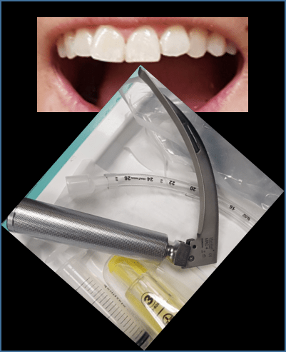
An intubation manikin was modified to contain concealed strain gauges measuring axial and lateral forces applied to each of the four maxillary incisors.
104 consultant and trainee anaesthetists in the two hospitals were invited to intubate the manikin, each using three different laryngoscopes and in two different airway conditions (normal and difficult). The purpose of the study was not revealed to the participants.
The axial and lateral forces exerted on each of the four maxillary incisors was recorded during each intubation.
A size 3 Macintosh blade and a KingVision standard aBlade size 3 were used in both hospitals, while due to differences in local familiarity a GlideScope AVL size 3 was used as the third laryngoscope in one hospital and a C-MAC Macintosh blade size 3 was used in the other.
Participants completed an anonymous questionnaire recording gender, level of training and experience of relevant laryngoscopy techniques.
Primary Outcome
The peak force on the maxillary incisors during endotracheal intubation with different laryngoscopy techniques and in normal and difficult airway conditions was the primary outcome.
Secondary Outcome
The impact of operator gender and level of training were analysed as secondary outcomes.
Statistics
Each of the three videolaryngoscopes was compared to direct laryngoscopy, which served as a common reference group. The Chi-square test was used for categorical variables and paired non-parametric analysis with the Wilcoxon signed-rank test was used to compare continuous variables of dependent samples.
Results
When compared to direct laryngoscopy, the hyperangulated videolaryngoscopes (GlideScope & KingVision) caused significantly lower peak forces on the maxillary incisors in both normal and difficult airway conditions.
Difficult airway conditions resulted in significantly higher peak forces than normal airway conditions when the C-MAC or Macintosh blade were used, but not when the hyperangulated videolaryngoscopes were used.
The left lateral incisor received the greatest force during all techniques and in both normal and difficult airway conditions with two exceptions; use of the GlideScope in normal conditions placed greatest force on the left medial incisor, and use of the KingVision in difficult conditions placed most strain on the right lateral incisor.
Gender was not associated with significant differences in peak forces, though consultant anaesthetists did generate higher peak forces than trainees when intubating with a direct laryngoscope in normal airway conditions and with a C-MAC in all conditions.
Conclusions/Discussions
Forces exerted by the Macintosh and GlideScope were comparable with those in other trials, but those recorded for the C-MAC were higher (more than double in normal airway conditions). Lack of blinding in those studies is suggested as a possible reason for this. Another is the relative lack of experience using the C-MAC in the anaesthetists using it in this study. Poorer Cormack-Lehane views were achieved in this study than in comparable trials which may indicate the manikin was difficult to intubate with this laryngoscope compared to others and real patients, requiring the use of greater force.
The authors suggest that the force applied to the right lateral incisor may be due to levering forces by the rigid tracheal tube stylet.
The higher forces generated by consultants using direct laryngoscopes in this study were not easily explained, but greater hesitancy and caution among trainees was suggested by the authors as a potential cause.
The authors conclude that the indications for the use of a hyperangulated videolaryngoscope may be extended to include patients at increased risk of dental damage, regardless of their anticipated difficulty of intubation.
Stated Limitations from the Study
The use of even a high-quality manikin cannot provide an exact replication of the intubating mechanics of a real patient. Likewise, the simulation of difficult airway conditions in this study only provides one of numerous possible scenarios where intubation is complicated.
As the study was powered based on the primary outcome, where the anticipated effect size was quite large, it is possible that it was under-powered to detect a difference due to gender of operator.
The two departments used different videolaryngoscopes, so comparison is more prone to confounding factors.
Discussion from Journal Club Meeting
If blinded to the purpose of the study, would participants have taken less care with a manikin’s teeth than a real patient’s.
Some already have a low threshold for intubating with a hyperangulated videolaryngoscope, and this adds weight to doing so in cases where the patient’s teeth are more vulnerable.
Summary by Dr D Baily. Journal Club Meeting 28 November 2019.
